What happened
According to a paper published in PLOS Digital Health, researchers tested whether a retinal biological age score, RetiAGE, derived from AI analysis of retinal photographs, is associated with skeletal health and future osteoporosis. The team evaluated cross-sectional associations in the Singapore PIONEER cohort (n = 1,965) that had same-day retinal images and DEXA bone-mineral-density (BMD) measurements, and tested prospective prediction in a UK Biobank sample (n = 43,938) without osteoporosis at baseline, followed for approximately 12.2 years (PLOS Digital Health). The paper reports that higher RetiAGE correlated with lower BMD in hip and femoral regions and higher FRAX-derived fracture-risk scores in PIONEER after multivariable adjustment (PLOS Digital Health). In UK Biobank, RetiAGE predicted incident osteoporosis with an adjusted hazard ratio of 1.12 per standard deviation and a dose-response pattern across quartiles; participants in the highest quartile had HR 1.40 versus the lowest quartile (PLOS Digital Health). The authors also report that adding RetiAGE to an osteoporosis self-assessment model increased the C-index from 0.585 to 0.635 (PLOS Digital Health).
Technical details
Editorial analysis - technical context: The RetiAGE algorithm was trained to estimate retinal biological age as a probability-like score and was applied to two large cohorts, enabling both cross-sectional and longitudinal analyses. The paper describes multivariable models adjusted for standard osteoporosis risk factors including:
- •age, sex, calcium intake, diabetes, hypertension, smoking, physical activity, and glucocorticoid use (PLOS Digital Health).
The reported effect sizes are modest but consistent: per-SD hazard ratio 1.12 is small at the individual level but the highest quartile versus lowest quartile HR 1.40 suggests the signal concentrates in the tails. The improvement in discrimination reported by the authors, a C-index increase of 0.050, is measurable but remains in a range that is not yet sufficient for clinical decision-making on its own (PLOS Digital Health).
Context and significance
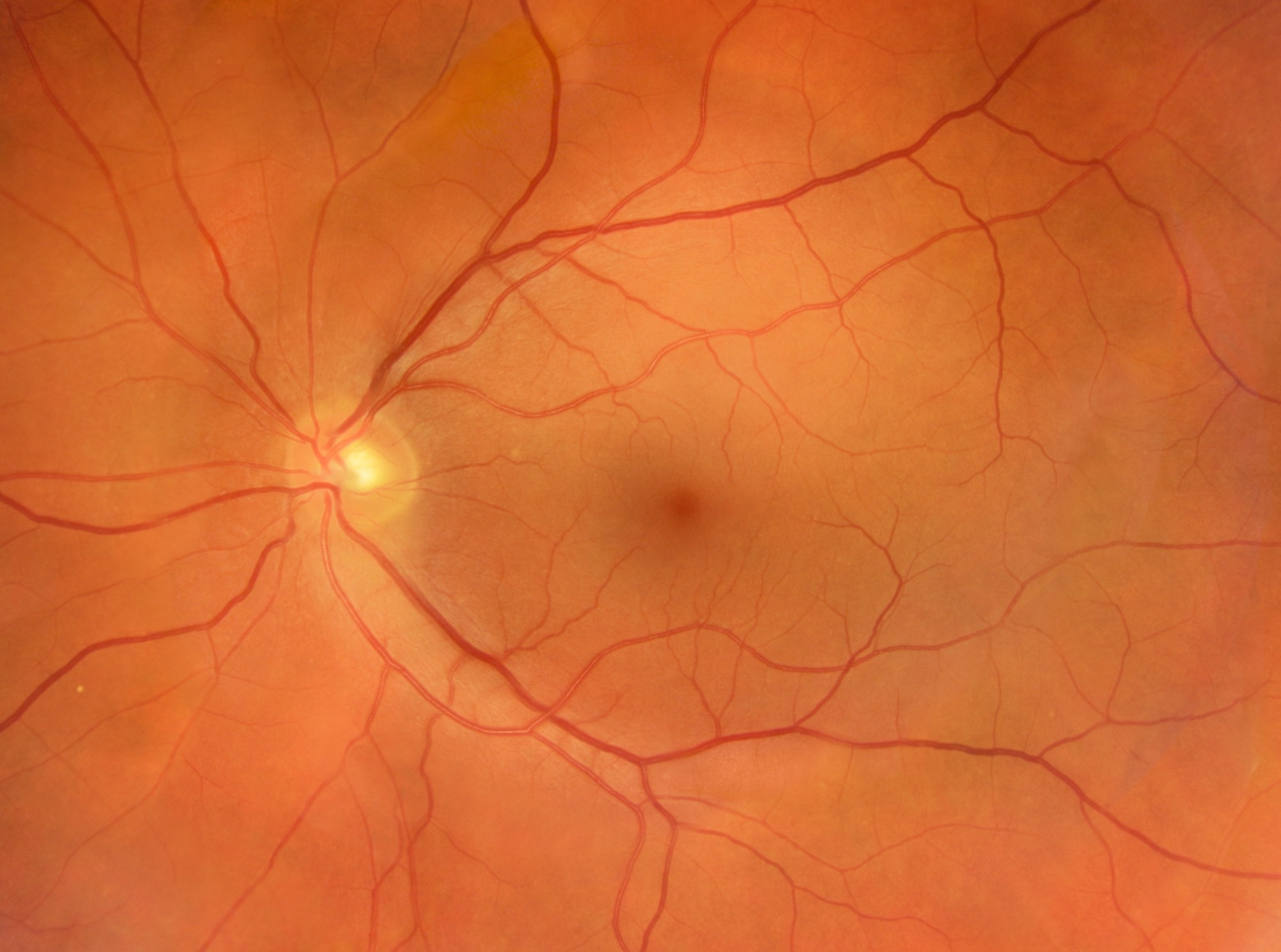
This study fits into a broader research trajectory where retinal phenotypes are used as accessible, noninvasive proxies for systemic vascular, metabolic, and inflammatory aging processes. Prior work has linked retinal features to cardiovascular risk, cognitive decline, and kidney disease; this paper extends that line to skeletal health using large population cohorts (PLOS Digital Health; Discover Magazine; ScienceAlert). For practitioners, retinal imaging is attractive because fundus photography is low-cost, widely available in eye care settings, and does not require ionizing radiation, unlike DEXA.
Observed limitations documented in the paper and covered in reporting include cohort composition and effect size. The PIONEER cohort provides contemporaneous retinal and DEXA data but is cross-sectional, limiting causal inference (PLOS Digital Health). The UK Biobank analysis adds longitudinal evidence but depends on osteoporosis ascertainment from health records and imaging substudies. The authors report adjustments for multiple confounders, but residual confounding and the generalizability beyond the studied populations remain open questions (PLOS Digital Health; News-Medical).
What to watch
For practitioners: follow-up work to validate RetiAGE performance in independent clinical screening populations and to test whether retinal-derived risk stratification materially changes clinical pathways for osteoporosis screening. Key indicators include replication of effect sizes in diverse ancestries, demonstration of incremental predictive value for fracture outcomes rather than diagnosis codes, and cost-effectiveness analyses comparing opportunistic retinal screening plus targeted DEXA against usual care. Researchers and implementers should also examine model calibration across age and sex subgroups and the potential for confounding by ocular disease and imaging quality.
Editorial analysis: If replicated, retinal-aging biomarkers could become a low-cost tool for population-level risk stratification, shifting some opportunistic screening to vision-care settings. However, current reported discrimination gains are modest and require prospective implementation studies before clinical adoption.
Key Points
- 1RetiAGE associates with lower bone mineral density and higher fracture-risk scores, across Singapore and UK cohorts (PLOS Digital Health).
- 2Adding RetiAGE to a self-assessment model increased the C-index from 0.585 to 0.635, a measurable but modest improvement (PLOS Digital Health).
- 3Industry context: retinal imaging is a scalable, noninvasive modality for systemic aging biomarkers, but replication and clinical validation remain necessary.
Scoring Rationale
This is a notable research result linking an AI retinal-age biomarker to osteoporosis risk across two large cohorts, offering a plausible pathway for low-cost opportunistic screening. The effect sizes and discrimination gains are modest, so practical impact depends on replication and prospective validation.
Practice with real Health & Insurance data
90 SQL & Python problems · 15 industry datasets
250 free problems · No credit card
See all Health & Insurance problems