Researchers review AI methods for meningioma MRI detection
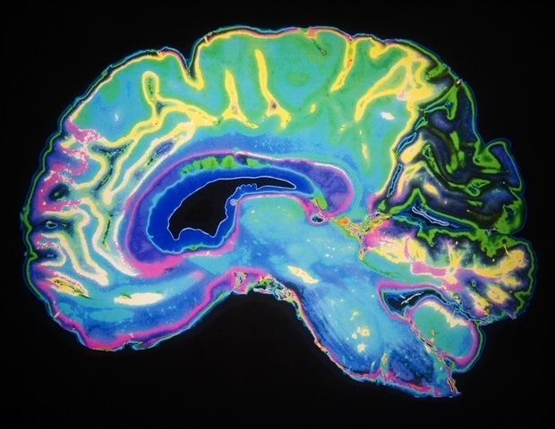
News-Medical reports that researchers from the University of Auckland, Auckland City Hospital, and Matai Medical Research Institute reviewed 34 articles on AI-based meningioma delineation published from 2020 to 2025. The review appears in the journal Brain Network Disorders (available online February 13, 2026, Volume 2, Issue 1, March 26, 2026), News-Medical reports. Lead author Dr. Hamid Abbasi is quoted: "More advanced AI models have clearly contributed to improved tumor delineations over time, with their performances becoming better and more consistent." News-Medical reports that the review found that "smarter models matter more than additional data or better scans," and that studies used public repositories such as Figshare and BraTS alongside many custom hospital datasets. The article also reports that contrast-enhanced T1 MRI was the most effective and commonly employed imaging method, and that model performance was commonly evaluated using the Dice score metric.
What happened
News-Medical reports that a team from the University of Auckland, Auckland City Hospital, and Matai Medical Research Institute conducted a systematic review of 34 studies on automated meningioma delineation from MRI, covering publications from 2020 to 2025. The review is published in Brain Network Disorders (online February 13, 2026; Volume 2, Issue 1, March 26, 2026), News-Medical reports. The article quotes lead author Dr. Hamid Abbasi: "More advanced AI models have clearly contributed to improved tumor delineations over time, with their performances becoming better and more consistent," and reports his statement that "smarter models matter more than additional data or better scans."
Technical details
News-Medical reports that the reviewed studies used a mix of public repositories, including Figshare and BraTS, and many institution-specific datasets. The coverage highlights contrast-enhanced T1 MRI as the most commonly used and effective modality for meningioma detection. The article also notes that segmentation performance is frequently reported using the Dice score, a metric that quantifies spatial overlap between predicted and reference segmentations.
Editorial analysis - technical context
Industry-pattern observations: improvements in model architecture often yield larger gains in segmentation quality than proportional increases in dataset size, according to the review findings cited by News-Medical. For practitioners, this aligns with broader experience in medical-image segmentation where architectural choices, inductive biases, and loss-function design can materially affect small-lesion and boundary delineation performance independent of training-set scale.
Context and significance
Industry context: automated meningioma segmentation affects diagnostic workflow speed, inter-reader consistency, and surgical and radiotherapy planning. The review's emphasis on architecture over raw data volume underscores a practical trade-off for research groups and vendors working under typical clinical constraints, such as limited labeled cases and scanner heterogeneity.
What to watch
For practitioners: monitor whether future studies from the field publish external multi-center validations, open-source model code, or prospective clinical evaluations that report Dice and other robustness metrics across scanner types. Observers should also track efforts to standardize dataset curation (contrast protocols, annotation protocols) and benchmarks that reflect small and irregular tumor segmentation challenges.
Key Points
- 1Review of 34 studies finds model architecture improvements drove segmentation gains more than larger datasets, highlighting design over scale.
- 2Contrast-enhanced T1 MRI is the most used modality for meningioma detection, so modality-specific preprocessing remains important for comparability.
- 3Practitioner-critical metrics like the Dice score dominate reporting; external multi-center validation and robust benchmarks remain key next steps.
Scoring Rationale
This literature review consolidates evidence that architectural advances produce measurable gains in meningioma segmentation, a notable finding for medical-imaging practitioners and researchers. The story is important but domain-specific, so it rates as a notable update rather than a field-wide paradigm shift.
Sources
Public references used for this report.
Practice with real Health & Insurance data
90 SQL & Python problems · 15 industry datasets
250 free problems · No credit card
See all Health & Insurance problems
